Microscope Facilities
Leica Stellaris 8 White Light Laser Scanning Confocal (w/ FLIM and FCS)
Stand - Leica DMi8 automated microscope
Objectives - 10X NA 0.4, 20X NA 0.75, 25X NA 0.95 NA (water), 40X NA 1.4 (oil), 63X NA 1.4 (oil), 63X NA 1.2 (water)
Stage - Encoded, closed loop automated stage with autofocus and Piezo Z axis
Illumination - 80 MHz pulsed white light laser tunable in 1 nm increments from 440-790 nm with 8 lines available simultaneously; 405 nm continuous laser
Detector - 5 HyD detectors with spectrometer for spectral scanning from 410-850 nm
Confocal - Stellaris8 resonance scanning confocal scan head
Super-resolution - Lightning deconvolution based super-resolution to 150 nm
Time-correlated photon counting - Enables fluorescence lifetime measurements and fluorescence cross correlation spectroscopy
Computer - HP, Intel Xeon Gold w/ 8 cores, 192 GB RAM, NVIDIA GPU w/ 24 GB RAM
Software - LasX with Tau-sense, Phasors, Lightning, 3D Visualization
Primary Uses - Optical sectioning confocal, FLIM, FCCS, Super-resolution, FRAP, DIC
Purchased in 2020 with funding from NSF (Nick Galati, PI; Lynn Pilitteri, Shawn Arrelano, Jeanine Amahcer, Jackie Rose, co-PI; NSF MRI 2019228)
LEICA STELLARIS 8
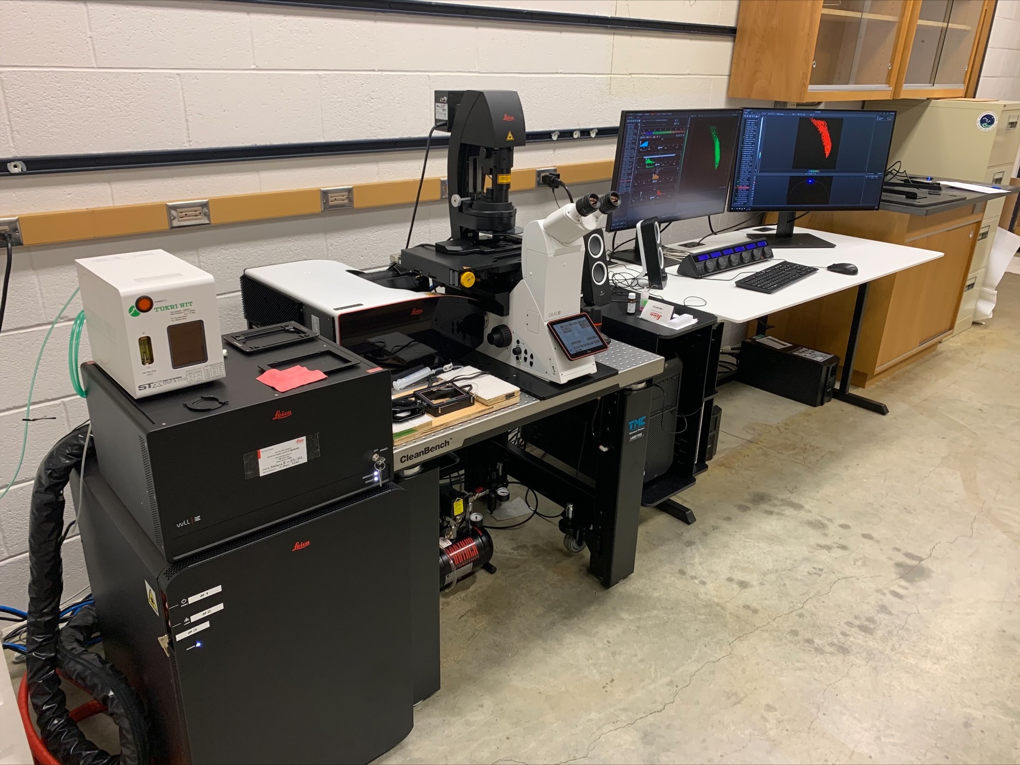
The Leica Stellaris 8 is the most modern and fully featured laser scanning confocal microscope offered by Leica. It is a workhorse microscope that enables single molecule imaging all the way up to whole embryo imaging. The microscope is managed by the SciTechServices core facility and maintained through a full service contract with Leica Microsystems. Train with Nick Galati.
DeltaVision with Olympus IX70 base
Room: Biol 452
Objectives: 20X air UPlan Apo NA 0.75; 40X air UPlan Apo NA 0.80; 60X oil UPlan Apo NA 1.4 (oil immersion); 60X Plan Apo NA 1.4 (water immersion).
Fluorescence filter sets: DAPI, Fluorescein, Rhodamine, Texas Red, CY5, GFP, CFP, YFP, mCherry
Camera: Photometrics HQ2 Cool Snap
Fluorescence illumination: Xenon lamp
Microscope capabilities: Fluorescence, DIC, Deconvolution microscopy, 3-D reconstructions
Purchased in 1998 and upgraded in 2009 with funding from NSF
(David Leaf, PI); and WWU STF (David Leaf, PI)
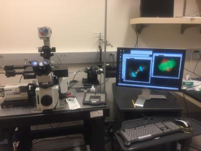
The Deltavision is useful for 3-D reconstructions of computationally deblurred fluorescence specimens and live cell imaging. To be approved for using the Deltavision, users should have experience with fluorescence microscopy and train with Nick Galati.
Leica iDM6000 inverted automated microscope
Room: Biol 455C
Objectives: 10X HCX Pl Fl NA 0.3, 20X HCX Pl Fl NA 0.4, 40X HCX Pl Fl NA 0.60; 60X HC Pl Apo NA 1.4 (oil immersion), 100X HCX Pl Fl NA 1.25 (oil immersion)
Fluorescence filter sets: DAPI, Fluorescein, Rhodamine, GFP, CFP, YFP, CY5, mCherry
Camera: Leica DFC3000G cooled CCD
Fluorescence illumination: Mercury Metal Halide
Microscope capabilities: Fluorescence, DIC, phase contrast
Purchased in 2014 with monies from WWU CSE and friends and alumni of Biology (Joann Otto - Fundraiser).
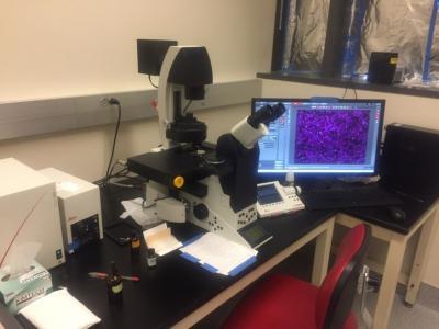
The Leica iDM6000 has all of the capabilities of the Leica DMRB but because it is fully automated, it is easier to image with than the manual microscope. To be approved for using the Leica iDM6000, users should have some familiarity with optical microscopy and train with Nick Galati.
Leica DMRB microscope
Room: Biol 452
Objectives: 10X Fluotar NA 0.3, 20X Fluotar air NA 0.5, 40X Fluotar NA 0.70; 40X N-plan NA 0.65, 100X N plan NA 1.25 (oil immersion)
Fluorescence filter sets: DAPI, Fluorescein, Rhodamine
Camera: Spot RT3 cooled CCD with color filter option
Fluorescence illumination: Mercury Metal Halide
Microscope capabilities: Fluorescence, DIC, phase contrast
Purchased in 1994 with funding from NSF (David Leaf, PI) and WWU STF (David Leaf, PI).
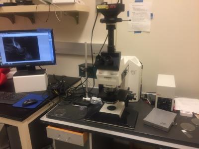
The Leica DMRB is an all around research-grade manual microscope. To be approved for using the Leica DMRB, users should have some familiarity with optical microscopy and train with Nick Galati.
Olympus BX60 upright microscope
Room: Biol 455C
Objectives: 10X Achromat l NA 0.25, 20X UPlan Fl NA 0.5, 40X UPl Fl NA 0.75; 100X UPl Fl NA 1.3 (oil immersion)
Fluorescence filter sets: DAPI, Fluorescein, Rhodamine
Camera: SPOT RT3 cooled CCD
Fluorescence illumination: Olympus UGLGPS Mercury Metal Halide
Microscope capabilities: Fluorescence, DIC, phase contrast
Purchased in 1996 with monies from Biology Building Equipment Funds and WWU STF (David Leaf, PI)
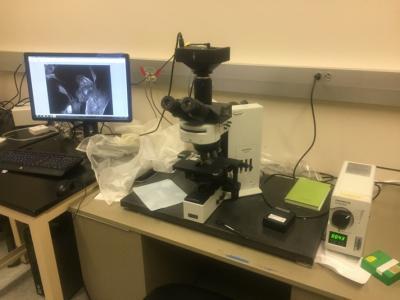
The Olympus BX60 is the most basic research-grade microscope and is simpler to use because it lacks DIC optics. To be approved for using the Leica DMRB, users should have some familiarity with optical microscopy and train with Nick Galati.
Imaris Workstations:
The Biology Department has two Imaris workstations for rendering and analyzing fluorescence images.
CellAsic Omix Microfluidic Platform:
This platform enables control of temperature, CO2, and perfusion of media into 4 imaging chambers for live cell imaging.